The University of Manchester Faculty of Life Science has a Polara transmission electron microscope for high-resolution cryo-electron microscopy. The microscope was funded by generous grants from the Wellcome Trust and the University of Manchester.
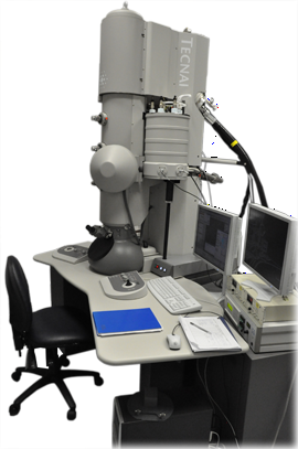
The Tecnai G2 Polara is fitted with a Gatan post column energy filter. This microscope is a cryo-transmission EM with a 300 kV field emission gun (FEG), which allows high resolution imaging of proteins in their native state.
Current research includes:
- Defining the molecular basis of cell-mediated ECM assembly.
- Elucidating the molecular basis of cell-matrix adhesion and signalling.
- Determining the structure of ECM macromolecules.
- Determining the structure and function of P2X receptors and the cystic fibrosis transmembrane conductance regulator (CFTR) protein.
- Examining structural variation in viral coat proteins
To complete these studies the microscope is being used to generate thick section tomograms of tissue sections and cryo-single particle reconstructions of macromolecules frozen in vitreous ice.
-
User
Access
-
Single
Particle
-
Electron
Tomography
-
Ancillary
Equipment
Our electron microscope facility is available to both internal and external users for independent or collaborative projects that fit the expertise of our members.
If you would like to apply to use the Polara microscope then please complete and return the following form: Contact form
If you wish to discuss the potential benefits for your current research then you can contact
Dr Tobias Starborg:
tel: +44(0)1612755170 or
email: 
The Polara can be loaded with 6 samples, for convenient screening of multiple samples. The microscope has reliable grid loading and specimen transfer (achieved through a protected environment in the loading bay, and the transfer system under vacuum).
The microscope stage is stable to allow high resolution imaging. The design also gives a low rate of specimen contamination in the microscope.
To collect high resolution data the images can be captured on film or the 4k by 4k Gatan USC 4000.
Electron tomography is a method of generating a 3-dimensional reconstruction of a thick section by collecting a series of tilted images. The Polara is equipped with tilt rotate cartridges that allow high angle (+-70 deg) tilt and 90 deg rotation of the grid without removing the sample from the microscope vacuum. The use of a cartridge-based system also increases the stability of the sample within the beam and reduces errors in stage movement during data acquisition.
The Polara at Manchester has the following tomography imaging options:
- Gatan Ultrascan USC 4000 (4k x 4k CCD)
- Gatan Imaging Filter 2002 (2K by 2K GIF)
The following software is installed to allow collection of data sets and generation of tomograms:
The University of Manchester EM facility has a large selection of cryo-EM equipment to facilitate user sample preparation including:
- Baltec HPM 010
- Leica EM-PACT
- Leica EM AFS2 freeze substitution
- Leica EM UC6 cryo microtome
- FEI Vitrobot
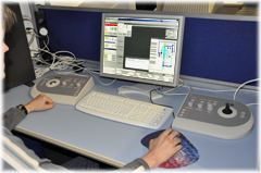
The FEI G2 Polara microscope is housed in a purpose built room within the electron microscope facility. The air handling maintains the temperature to within one degree centigrade. The room is well insulated from noise and vibrations. It is also possible to use remote access to control all of the microscope functions. This means that the Polara is able to reproducibly collect high resolution data.
We are happy to give advice about installation and running the facility for people who are planning on installing a Polara (or similar) microscope.